
Dental CT Scan
Medically reviewed by Dr. Kayla Lebeau, DDS
Seeing the jaw in three dimensions
An ordinary dental X-ray is a flat picture of a solid object. Everything is layered on top of everything else, and depth simply does not exist in the image. For most everyday dentistry that is perfectly adequate.
For anything involving surgery, bone or root anatomy, it is not. A CBCT scan, cone beam computed tomography, captures the jaw as a three-dimensional volume that can be sliced and measured in any direction. Bone height and width to the millimeter, the exact path of the nerve in the lower jaw, the floor of the sinus, and root canals that a flat image cannot separate.
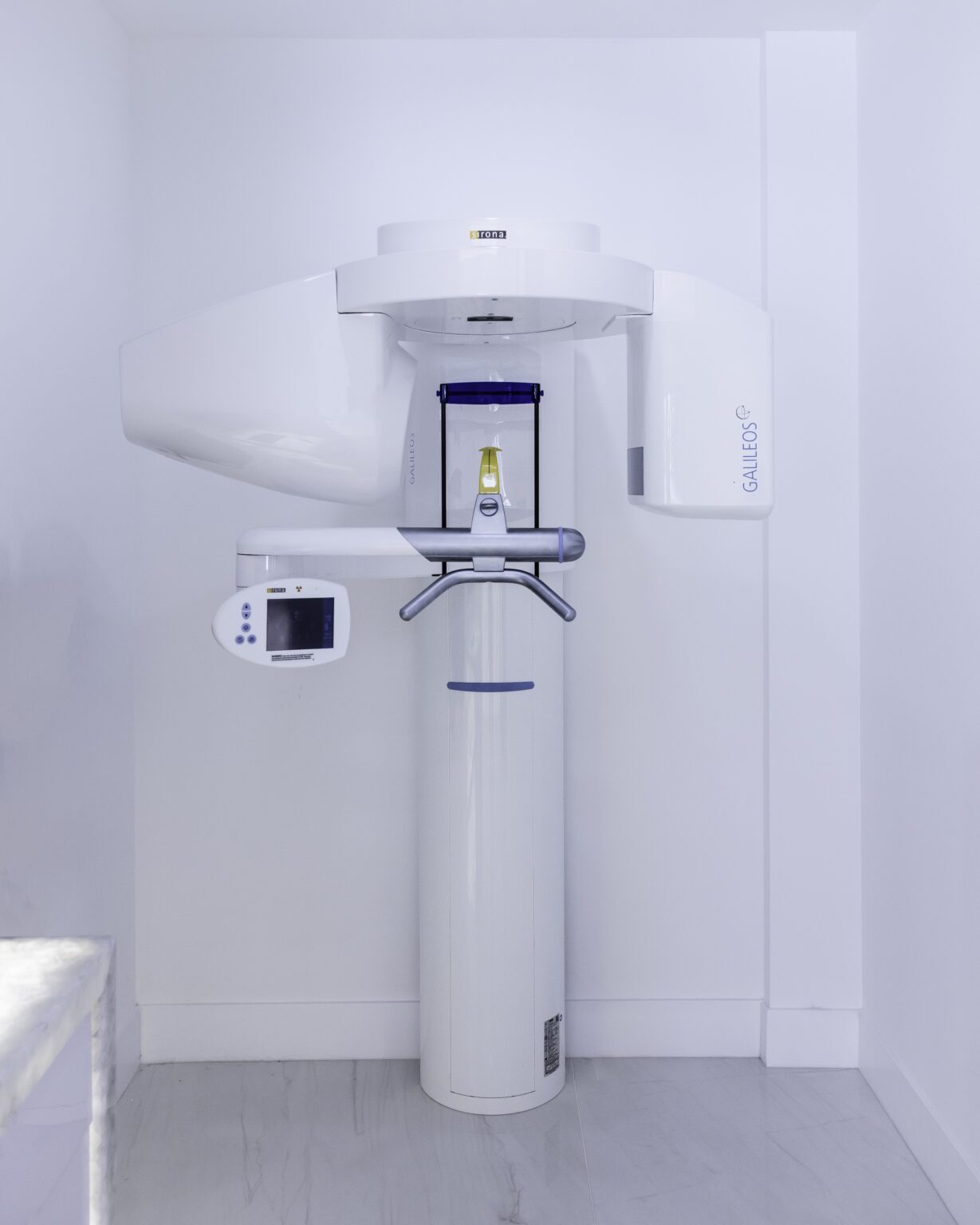
We have the scanner in the practice, so the scan happens during your appointment rather than at a referral across town.
- TakesUnder a minute in the machine
- ComfortNothing enters your mouth
- RadiationFar lower than a medical CT
- ResultsReviewed with you on screen
A 3D scan is genuinely useful when
- Planning dental implants. Bone volume and nerve position decide what is possible
- Assessing whether a bone graft or sinus lift is needed
- A wisdom tooth sits close to the nerve in the lower jaw
- A root canal has failed and a canal may have been missed
- Investigating a suspected crack, a cyst, or persistent infection
- Assessing the jaw joint, or an impacted tooth in a child
What happens, and the honest position on radiation
- PreparationYou remove earrings, glasses and anything metal around the head. Nothing goes in your mouth and nothing touches you.
- The scanYou rest your chin on a support and stay still while the arm rotates once around your head. Under a minute, usually a good deal less. There is no tunnel and nothing encloses you. People who find medical CT or MRI difficult are generally fine with this.
- ReconstructionThe software builds the 3D volume from the images.
- Reviewing it with youWe look through it on screen together. Seeing your own jaw, the nerve and the bone makes the plan considerably easier to follow than being told about it.
- PlanningFor implants, positions are planned virtually on the scan before surgery. That planning is what turns an implant placement from a judgment into a measurement.
On the dose: a CBCT scan uses more radiation than a single dental X-ray and far less than a medical CT of the head. The field of view is limited to the area we need, which keeps the dose to a minimum. We take one when the information will change what we do, not routinely, and not because the machine is there.

What it changes in practice
- Implants become planned rather than estimated. Exact bone width, height and density, and the position of the nerve, are known before surgery instead of discovered during it.
- Grafting decisions become clear. Whether you need a graft or a sinus lift is a measurement, not an opinion.
- Missed root canals are found. The commonest cause of a failed root canal is a canal that never appeared on a flat X-ray.
- Wisdom tooth surgery gets safer. Knowing precisely where the nerve runs relative to the roots is what protects the feeling in your lip.
- Cracks and cysts are located that flat imaging genuinely cannot show.
Is it safe?
Yes, within the normal principles of medical imaging. The dose is low, the field is limited to the area of interest, and we only take one when it will change the treatment.
Is it claustrophobic?
No. Nothing encloses your head, the arm rotates around you in the open, and it is over in well under a minute.
Does it hurt or involve anything in my mouth?
Neither. You sit still and that is the whole procedure.
Can I get a copy of my scan?
Yes. If you are seeing a specialist elsewhere we can provide the data file so it does not need repeating.
Will my insurance cover it?
Coverage varies by plan and by why it is being taken. We will check and tell you the position beforehand.
Can I have one if I am pregnant?
We avoid elective imaging in pregnancy and postpone unless it is urgent. Tell us if you are or might be pregnant.
Ask whether a 3D scan would help your case
- (310) 860-6969Someone here answers.
- Request a visitWe will call to confirm.
- Pick a time onlineChoose your own slot.
What a 3D scan shows that an X-ray cannot
A normal dental X-ray is a flat shadow of a three-dimensional object. Structures overlap, and depth is invisible. A cone beam CT reconstructs the anatomy in three dimensions, so it can be viewed from any angle and measured accurately.
That matters in specific situations:
- Implant planning. Measuring bone width and height, and the exact distance to the nerve canal or sinus floor. This is the main reason we own one.
- Impacted wisdom teeth. Showing the true relationship between the roots and the nerve before removal.
- Root canal problems. Finding an extra canal or an infection at a root tip that a flat film hides.
- Unexplained pain, fractures, and assessing the jaw joint.
The scan itself takes under a minute. You stand or sit still while the machine rotates once around your head. Nothing touches you and there is nothing to hold in your mouth.

Radiation, honestly
A dental cone beam CT uses more radiation than a standard dental X-ray and substantially less than a medical CT scan of the head. The dose varies with the size of the area scanned, and a small focused scan of one region is a fraction of a full-jaw one.
For context, the dose from a typical dental CBCT is broadly comparable to a few days to a few weeks of ordinary background radiation from the environment. That is a real exposure, not a trivial one, and it is why we do not scan routinely or as part of a check-up.
We take one when it will change the treatment decision, and we can tell you in advance what question it is meant to answer. If we cannot explain that, you should not have the scan.
We use the smallest field of view that answers the question, and a thyroid collar where appropriate. Tell us if you are or might be pregnant.